Located in the Department of Chemistry & Biochemistry room 4608 PSB-N, the X-ray lab has the
instrumentation to perform small-molecule single-crystal and
powder-diffraction experiments. X-ray diffraction is a
nondestructive technique widely used in physics, chemistry,
biology and materials science to characterize the structures and
properties of solid state materials. Single-crystal
diffraction can provide precise information about 3-D molecular and
crystal structures, while powder diffraction is mainly
used in qualitative and quantitative phase analysis, the
characterization of various properties such as particle size,
strain, and stress of polycrystalline samples.
The PC-controlled, fully automated instruments come with
well-developed analytical software packages giving users the
capability for comprehensive solutions from the acquired
data. The facilities provide access for both on-campus and
off-campus users. Lab users can either conduct the experiments
themselves after operation and safety training or submit the
samples to lab staff who will provide the service of data
collection and processing. To
schedule an experiment, users
should contact the lab staff. Periodically, the X-ray lab staff
organizes group training in instrument operation and data
analysis. Individual training is also available upon request.
|
Rigaku Synergy Diffractometer
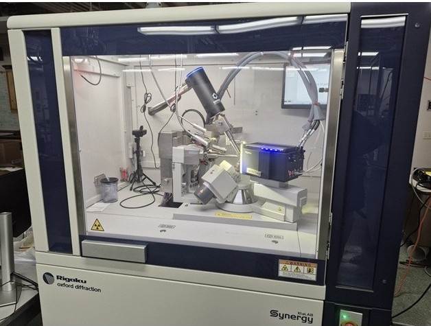 |
- Chemical and Biological Crystallography
Rigaku Synergy XtaLab with photon count detector and duo micro-focus X-ray sources. It has very bright focused beams of 0.1mm diameter area at sample site, and can be switched between Mo and Cu X-ray sources seamlessly in 1 min. It allows to collect data from crystals as small as 10 micron. The photon count detector HyPix-Arc 100, with the latest technology to detect X-ray photon directly, has little noise(background), enabling decent data with long exposure if needed. The curved detector screen can cover as wide as 100 degree of 2theta area per frame, making data collection much more efficient. The system is equipped with an Oxford 1000 plus liquid nitrogen cryostream. The data collection can be operated between 80K and 500K, allowing us to work on air/moisture sensitive samples, explore the temperature dependent phase transition and other investigation. With Cu X-ray source, it is also able to run screening of protein crystals and determine absoulte structure without presence of heavy elements. The 4-axis diffractometer allows identification of crystal faces, oriented X-ray scan and other analyses.
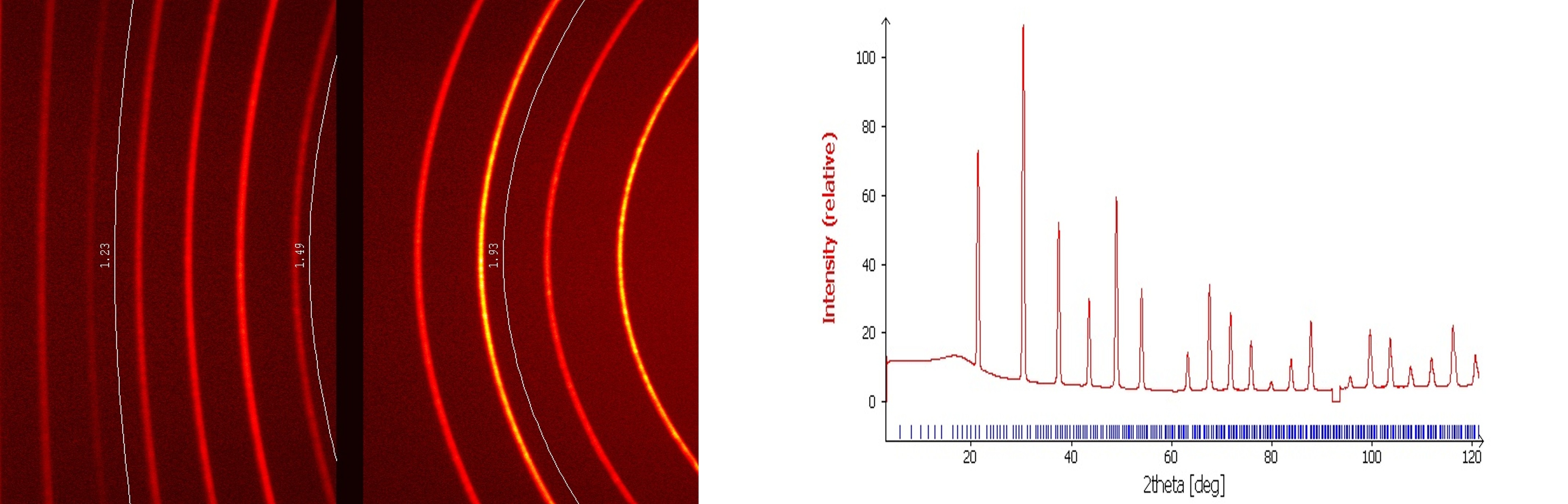
With the adjustable beam slits, the fine focused intense Mo and Cu X-ray can collecting high resolution 2D powder diffractions, providing an option to run speedy pxrd with a little amount of sample and multi X-ray sources.
|
|
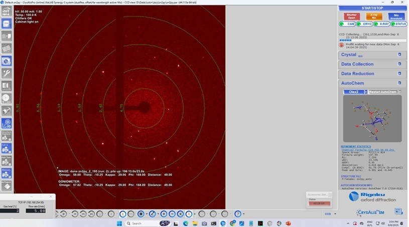
Single Crystal diffraction Data Collection and Real-Time Structure Determination(right)
 |
- Instrument Software
The instrument associated software package, CrysAliasPro, has very user friendly interfaces of indexing including twin indexing, data collection, data reduction and real-time structure detemination, automatioc CCDC database and local structure lattice search/match and other features. The lab also has OLEX and Shelx structure determination and refinement software packages.
|
|
2D Powder XRD from LB6 with Cu X-ray and Processed Spectrum
 |
|
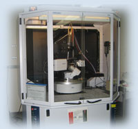
Bruker Kappa Apex II Diffractometer
|
- Bruker Kappa Apex II Diffractometer
The lab also has an operatable Bruker Kappa Apex II single-crystal diffraction system with CCD detector and 2Kw Mo fine focus sealed tube with TRIUMPH monochromator. The system is also equipped with Oxford 700 Plus Cryostream, which can perform temperature-dependent measurement from 80K to 500K. The instrument associates Bruker Apex 2 software package for data collection and process. It also has Shelx software installed for structure determination and refinements.
|
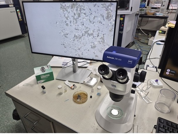
Zeiss Microscope
|
- Crystal Screening
The lab Zeiss Stemi 305 Microscope with Polarized Lens, High Resolution Digital Camera and monitors. The optical zoom is up to 64x, the high resolution image display on the monitor can help to pick the crystal(s), especially useful for demo and student training
|
XRD Structure and Property characterizations
|
 |
 |
 |
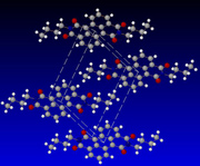
| Structure determination provides a vivid atomic connectivity and 3D molecular conformation and reveals the inter-molecular interaction and packing. |
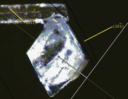
The indicies of crystal faces can be identified based on the orientation matrix. The diffractometer can rotate the defined face along a reference direction for visual observation or X-ray scan. |
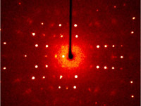
The oriented X-ray scan can help to observe defects, superlattice and other crystal properties. |
Lab Manager: Dr. Guang Wu
Office: 4610 Physical Sciences Building North
Phone: (805) 893-2399
Email:
wu@chem.ucsb.edu
|